Overview
Pediatric imaging is a subspecialty of radiology that provides a full range of imaging services for infants and children of all ages (up to age 18), including newborns. Imaging of the In utero developing fetus in pregnant women is also a part of this specialty. Our highly trained subspecialists use the latest in dedicated pediatric imaging equipment, including:
- Computed tomography
- Magnetic resonance imaging
- Ultrasound
- Fluoroscopy
Our specialists have completed years of additional training in pediatric radiology and pediatric neuroradiology to provide expertise in diagnosis and image-guided treatment. They received their fellowship training from some of the most highly respected programs in the country.
Pediatric imaging specialists perform patient studies at the VCU Health System, including various outpatient locations and the Children’s Hospital of Richmond (CHoR).
The latest equipment and technology provide non-invasive imaging studies and image-guided procedures, including:
- Computed tomography (CT) scan
- Uses low-dose radiation to create image “slices” of solid organs and systems
- EOS Imaging
- Produces 3D images using a highly sensitive detector that reduces radiation exposure
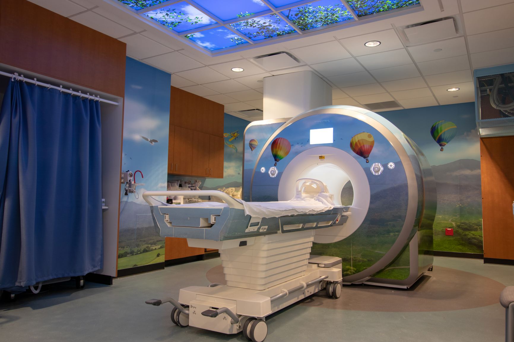
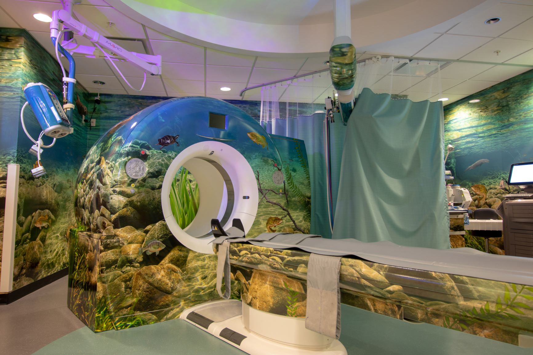
- Magnetic resonance imaging (MRI)
- Uses a strong magnetic field to create images of the body, and is best for seeing inside of soft organs
- Fluoroscopy
- Allows for live vision inside the body, and is often used during medical procedures
- Ultrasound (Sonography)
- Uses sound waves to capture real-time images and structures of internal organs
Pediatric Imaging Safety
Some pediatric imaging studies, including computed tomography scans and fluoroscopy require the use of radiation. We incorporate and use the American College of Radiology (ACR) sanctioned “Image Gently” principles of using every possible means to decrease the level of radiation exposure in children from these studies. We use child-specific imaging protocols and specialized imaging software to enhance image quality while minimizing radiation doses. We monitor our pediatric computed tomography radiation doses and report to the ACR for quality assurance. Our radiation doses are at the lower end of reporting hospitals and similar to other larger, free-standing children’s hospitals.
Learn More About Our Safety Office
Pediatric Imaging Faculty

Eman Sabah Mahdi, M.D.
Assistant Professor
Eman Sabah Mahdi, M.D.
Assistant Professor
Radiology
Director, Pediatric Radiology Fellowship Program
Email: eman.mahdi@vcuhealth.org
Address/Location:
Children's Hospital of Richmond at VCU
Chakradhar Mishra, MBBS, M.D.
Assistant Professor
Chakradhar Mishra, MBBS, M.D.
Assistant Professor
Radiology
Email: chakradhar.mishra@vcuhealth.org
Address/Location:
Children's Hospital of Richmond at VCU

Kathryn Starkweather Jones, M.D.
Associate Clinical Professor
Kathryn Starkweather Jones, M.D.
Associate Clinical Professor
Radiology
Phone: 804-628-7588
Email: kathryn.jones@vcuhealth.org
Address/Location:
Children's Hospital of Richmond at VCU
Gregory Vorona, M.D.
Associate Professor
Gregory Vorona, M.D.
Associate Professor
Radiology
Phone: (804) 828-9842
Email: gregory.vorona@vcuhealth.org
Address/Location:
Children’s Hospital of Richmond at VCU
Collaboration Improves Child Protection at VCU Health
Pediatric Imaging Takes Multidisciplinary Approach in Medicine to Improve Child Protection
Richmond, VA - September 2023 - Child abuse is an emotional diagnosis. Emergency physicians train to recognize clinical signs of child abuse but they rely on the expertise of Pediatric Imaging physicians to identify imaging findings that can help to differentiate between accidental injury and inflicted trauma. In these cases, physicians rely on imaging studies that can include radiographs, CT, and MRI scans to identify and characterize both clinically suspected and clinically non-suspected injuries. At VCU Health, Pediatric Imaging specialists collaborate with the Child Protection team, and together they offer complete pediatric care to the most vulnerable patients. This multidisciplinary approach in pediatric medicine also brings about important clinical innovations.
The recent addition of an anthropomorphic infant phantom with simulated rib fractures modeled from real child abuse victims presented at VCU Health was a multi-year collaborative project involving Pediatric Imaging, Diagnostic Medical Physics, and Pediatrics and Emergency Medicine. Gregory Vorona, M.D., Pediatric Imaging, Robin Foster, M.D., Director of the Child Protection Team, and Pei-Jan Paul Lin, Ph.D., Chair of Division of Diagnostic Medical Physics, worked together to bring about a first for VCU Health.
 “When an infant is brought into the ER and there is suspicion of child abuse a pediatric radiologist plays a particularly important role in looking for signs of acute and healing injuries,” Dr. Vorona says. “Often we do a series of radiographs, which is particularly helpful when detecting injuries to the extremities. However, when it comes to ribs, identifying fractures is not as simple using radiographs because there are multiple overlapping structures that can make some injuries difficult or even impossible to see. This is when CT can be more helpful both because overlapping structures no longer obscure potential injuries but also because CT allows for the creation of a 3D reconstruction of the rib cage. The sensitivity of CT is higher to detect acute and healing fractures, so it is easier to identify these injuries, and to differentiate fractures from congenital abnormalities. Because CT imaging provides more information, we thought in some cases we could use it instead of radiographs. However, to make it a routine diagnostic tool we had to figure out a way to lower the radiation dose.”
Dr. Vorona and Dr. Foster collaboratively explored the possibility of safely using CT imaging to assess pediatric cases where there is a high suspicion of child abuse, which would only be feasible if the radiation dose was low. To determine the CT dosing protocols required expertise from the Division of Diagnostic Medical Physics and, eventually, a custom infant phantom.
Dr. Lin and his colleagues were instrumental in the planning and development of this phantom. Dr. Lin who has longstanding relationships with the scientists and engineers at Norfolk, Va.-based Computerized Imaging Reference Systems, Inc. (CIRS), now part of Sun Nuclear, a Mirion Medical Company. CIRS is a leader in tissue simulation for medical physics and radiation therapy to calibrate specific imaging systems and determine the protocols that support the lowest radiation dose while still obtaining high quality images. As Dr. Lin worked with CIRS, Dr. Vorona collected non-identifiable CT images from real child abuse cases presented at VCU Health. The data provided by Dr. Vorona and Dr. Lin helped develop an infant phantom that simulated new and healing rib fractures.
 Radiological Technologist, David Burt, B.S, (R) (CT) (ARRT) under supervision of Monica Ghita, Ph.D., subjected the custom phantom to numerous CT scans at different radiation doses. These phantom study images were then reviewed by other faculty within VCU’s Pediatric Imaging section, including Drs. Eman Mahdi, Chakradhar Mishra and Kathryn Jones, to validate and confirm that the fractures in the phantom could be identified on the CT images. They found that an ultra-low radiation dose CT protocol achieved the sensitivity needed to detect rib fractures in the pediatric population, at a radiation dose comparable to the four standard radiographic views of the chest usually obtained.
Dr. Lin adds, “We received our first anthropomorphic infant phantom specifically designed for child abuse cases with simulated rib fractures, we are able to minimize the radiation level while maintaining image quality. We exposed the phantom to numerous CT scanners within VCU Health System so that Dr. Vorona could accurately identify fractures at a very low radiation dose, independent of which CT scanner was employed.” The radiation dose is nearly equivalent to that of four radiographs, thus, CT imaging is another viable tool to evaluate and diagnosis rib injuries in pediatric patients.