Overview
Virtual colonography or computed tomography colonography is a noninvasive procedure that uses computed tomography scanning to determine the presence of colon polyps.
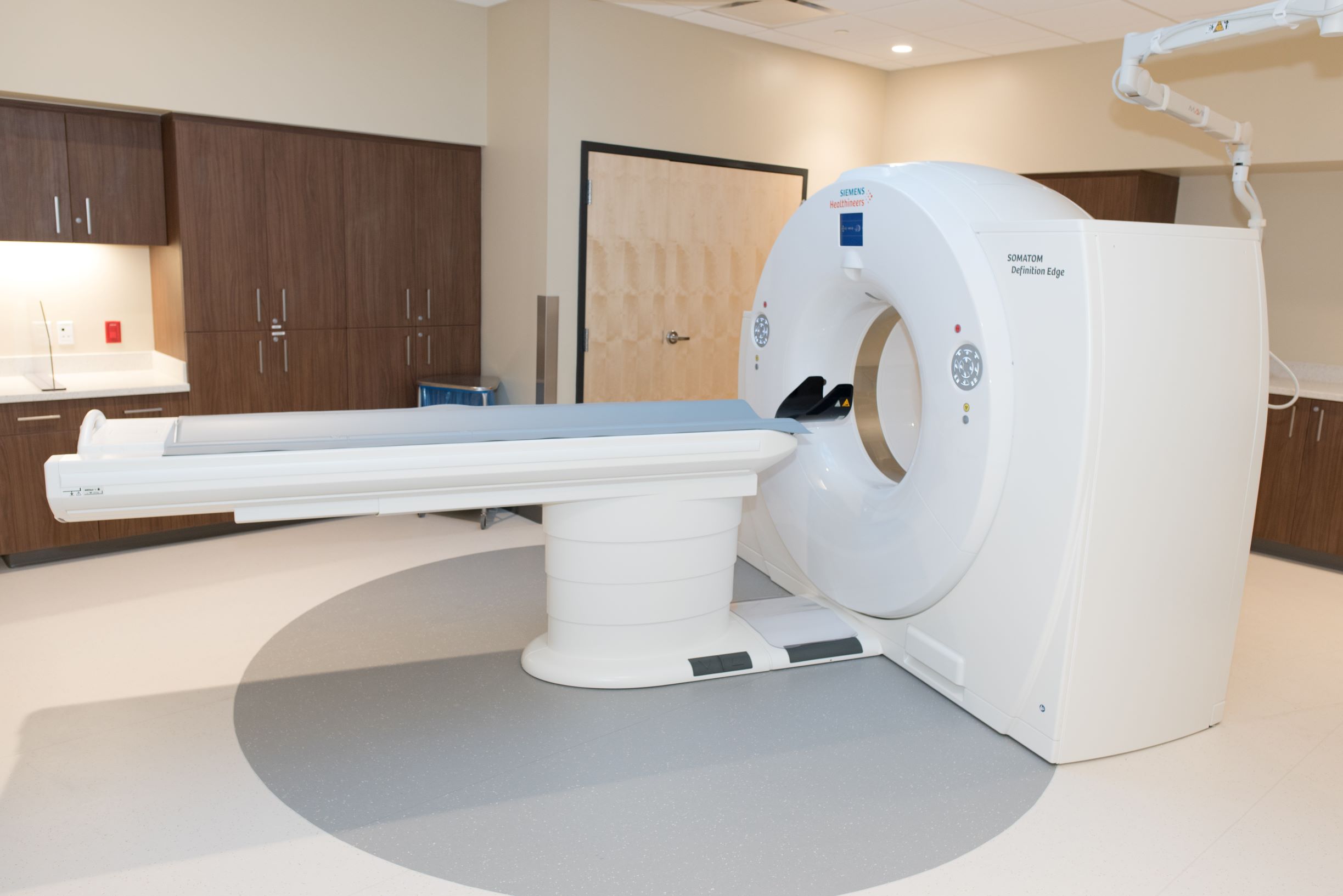
The VCU Department of Radiology was among the first in Virginia to test and use more patient-friendly methods of screening for colon polyps. Virtual colonography relies on specially developed computer software to recreate the inside of the patient’s colon using two-dimensional and three-dimensional images. The process allows a “fly-through” or “virtual” evaluation of the colon that provides the precise location and size of colon polyps.
Identification and removal of colon polyps before they become cancerous is the purpose of colon screening. Early studies show that virtual colonography is comparable to conventional colonography in terms of identifying polyps six millimeters or greater in size. Virtual colonography makes it possible to evaluate the entire colon, without risk of bowel perforation, or complications from sedation associated with standard colonography.
The VCU Department of Radiology uses virtual colonography as a diagnostic tool to confirm or rule out the possibility of cancer. If large polyps are identified, traditional colonoscopy is required for biopsy and removal.