Overview
The VCU Department of Radiology has specialty-trained radiologists who use magnetic resonance imaging of the breast to detect abnormalities of the breast and breast cancer.
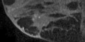
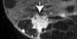
Magnetic resonance imaging of the breast (breast MRI) does not use ionizing radiation. Rather it uses a magnetic field to generate images of the breast before and after a contrast injection to detect cancer. The breast MRI is used as a supplemental procedure with mammography or ultrasound to evaluate abnormalities and screen high-risk patients for breast cancer.
Several risk assessment tools are used to estimate a woman’s breast cancer risk, including BRCAPRO, the Claus model and the Tyrer-Cuzick model. Based on different combinations of factors, these tools estimate breast cancer risk. Depending on the tool used, different risk estimates may be calculated for the same woman.
According to the American Cancer Society Guidelines, women at high risk should get a breast MRI and a mammogram beginning at age 30. Women at moderate risk should talk with their doctors about adding MRI to their yearly mammogram. Yearly breast MRI screening is not recommended for women whose lifetime risk of breast cancer is less than 15 percent.
The Breast Imaging Center at VCU Health is the first facility in Virginia to earn the American College of Radiology Breast Imaging Center of Excellence designation.
Patients and referring physicians visit the Breast Imaging Center at VCU Health for more information.